


Welcome to the November issue of The Surgeons’ Lounge. In this issue, Lisandro Montorfano, MD, a plastic surgery resident at Vanderbilt University Medical Center, in Nashville, interviews Dr. Huseyin Karagoz, MD, a plastic surgeon also at Vanderbilt University Medical Center. Dr. Karagoz provides up-to-date answers to the most common questions regarding the surgical management of lymphedema. We look forward to our readers’ questions and comments.
Also, the online version of this article will contain a special section titled “The History of Lymphedema Surgery—Past, Present and Future.”
Sincerely,
Samuel Szomstein, MD, FACS
Editor, The Surgeons’ Lounge
Szomsts@ccf.org
@YANKEEDOC44
The Surgical Management of Lymphedema

Lisandro Montorfano, MD: How would you define lymphedema and what are common contributors to this disease?
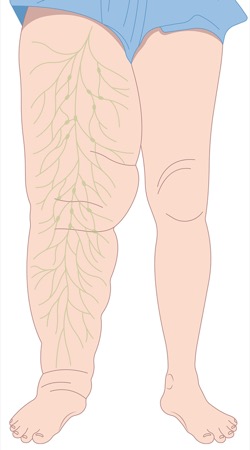
Huseyin Karagoz, MD: Lymphedema is a localized form of tissue swelling resulting from abnormal retention of proteinaceous lymphatic fluid within the interstitial compartment due to impaired lymphatic drainage or embryologic maldevelopment. One of the contributors is developmental lymphatic vascular anomalies, which are relatively rare. In developed countries, the most common contributors are cancer surgery and irradiation, while parasitic infection from Wuchereria bancrofti is the main contributor worldwide. Breast cancer survivors have an especially higher lymphedema risk than individuals with other types of cancer, due to the requirement of axillary lymph node removal as a part of the surgical treatment and irradiation. Any other surgeries that include lymph node removals such as vulvovaginal cancer or malignant melanoma are also risk factors for lymphedema development.
Dr. Montorfano: Could you expand on the most common lymphedema stagings? Do they correlate?

Dr. Karagoz: Staging systems for lymphedema are important in defining the severity of the problem, determining treatment modalities and predicting outcomes. Lymphedema can be staged based on clinical findings or confirmatory tests.
The most relevant and simple clinical staging system is the International Society of Lymphology (ISL) staging, although various other clinical staging systems have been proposed. ISL categorizes lymphedema into four levels of severity:
- Stage 0 (or Ia) refers to a latent or subclinical condition where swelling is not yet evident despite impaired lymph transport.
- Stage I represents an early accumulation of fluid relatively high in protein content which subsides with limb elevation, and pitting may occur.
- Stage II signifies that limb elevation alone rarely reduces the tissue swelling and pitting is manifest. Later in Stage II, the limb may not pit, as excess subcutaneous fat and fibrosis develop.
- Stage III encompasses lymphostatic elephantiasis, in which pitting can be absent and trophic skin changes such as acanthosis, alterations in skin character and thickness, further deposition of fat and fibrosis, and warty overgrowths have developed.
Conversely, various authors have also sought to stage lymphedema severity using diagnostic tests due to the need for better assessment of lymphatic vessels to be able to decide which type of surgical intervention is indicated. MD Anderson indocyanine green (ICG) staging system is frequently used for this purpose and categorizes the condition into four levels of severity:
- Stage I is defined by many patent lymphatic vessels with minimal dermal backflow.
- Stage II is defined by moderate patent lymphatic vessels and segmental dermal backflow.
- Stage III refers to few patent lymphatic vessels with extensive dermal backflow.
- Stage IV refers to no patent lymphatic vessels accompanied by severe dermal backflow.
Studies have shown that there is a weak correlation between ISL and ICG stages, indicating that physical examination and clinical staging are inadequate for assessing lymphatic function and deciding surgical intervention. ICG lymphography is required as a confirmatory test. It also should be noted that a limb may exhibit more than one stage, which may reflect alterations in different lymphatic territories.
Dr. Montorfano: What preoperative and intraoperative diagnostic modalities do you use to diagnose and classify this condition?
Dr. Karagoz: Although a presumptive diagnosis of lymphedema can be made with typical history and clinical exam, the diagnosis should always be confirmed with at least one of the two diagnostic tests—lymphoscintigraphy or ICG lymphography—because there are multiple other clinical conditions that may cause edema and swelling in extremities or any other body part. Furthermore, diagnostic tests can define the severity of the problem, determining the exact condition of lymphatic vessels, and can help predict outcomes, as I mentioned earlier.
Lymphoscintigraphy is a nuclear medicine imaging test that uses radioactive tracers and a gamma camera to detect lymphatic transport. It can define the patency of the lymphatics, the existence of backflow and any regional nodes. Lymphoscintigraphy is a useful preoperative diagnostic test for lymphedema and determining residual lymphatic function, but it is not sufficient in defining exact lymphatic vascular anatomy and establishing treatment preference.
ICG lymphography is a newer technique that has a higher sensitivity and specificity than lymphoscintigraphy. ICG lymphography allows for quick pathologic visualization of superficial lymph flow in real time, without radiation exposure. It precisely and reliably diagnoses, tracks and stages lymphedema severity, ranging from subclinical or early lymphedema to more advanced cases, and is more helpful to quantify the severity of the lymphatic obstruction and make the surgical plan. It can be used preoperatively as a diagnostic confirmatory test as well as intraoperatively for mapping lymphatic channels and guiding surgery.
Dr. Montorfano: Has surgery become the first-line treatment for this condition?
Dr. Karagoz: Not quite yet—complex decongestive physiotherapy is still the first-line treatment for the management of lymphedema. However, these modalities are all symptomatic and do not address real pathology. Surgery, on the other hand, directly addresses the main pathology of lymphedema and creates new paths for lymphatic fluid to be drained by bypassing obstructed or injured lymphatic vessels or nodes. However, surgery and decongestive physiotherapy should not be considered alternative modalities. Instead, they should be considered as two complementary treatment modalities to enhance their effectiveness. Having said that, surgical treatment of lymphedema has been significantly improved in recent years. The purpose is to provide a cure for lymphedema patients and when we look at the progress that we made so far, we can say that we are so close.
Dr. Montorfano: If the condition worsens and becomes difficult to manage, what surgical options do patients have?
Dr. Karagoz: Patients have immediate and late lymphatic reconstruction options. If a patient is in a high-risk group to develop lymphedema, such as a breast cancer patient who needs axillary lymph node dissection and radiotherapy, they do not have to wait until lymphedema development and worsens or becomes difficult to manage. We can perform immediate lymphatic reconstruction (originally called LYMPHA procedure) at the same time as the primary operation with surgical oncologists. We can find lymphatic vessels that have been already cut just after lymph node removal and create a bypass via lymph–venous anastomosis. This is a prophylactic surgery and the purpose is to repair lymphatic vessels immediately after injury.
As late lymphatic reconstruction, we have lymph–venous anastomosis (LVA) and vascularized lymph node transfer (VLNT) options. LVA is more beneficial at relatively early stages since the fluid component is dominant and it may be possible to drain the fluid by creating bypasses. VLNT has become the first treatment of choice in advanced stages. Having said that, technical refinements have improved the outcomes of LVA and have expanded its efficacy, creating an overlap between the indications for VLNT and LVA.
Other than these physiologic procedures, liposuction is a very helpful tool as a debulking procedure. We also can consider radical excision techniques if the condition is very severe and other surgical procedures are not convenient.
Dr. Montorfano: Is patient selection important at the time of performing these operations? Is there an ideal candidate?
Dr. Karagoz: Yes, indeed, patient selection is one of the most important factors for success. The initial evaluation should be comprehensive and include ICG lymphography to evaluate the status of lymphatic vessels and to make the correct surgery plan. Ideally, physiologic procedures should be performed as early as possible. Fluid will be predominant at earlier stages, while solid fat or fibrotic tissue components will be deposited at advanced disease. Physiologic procedures address the fluid component of the disease, so they would be more helpful for patients who have mild or moderate pitting edema. If a patient has significant solid edema, liposuction can be considered as initial surgical intervention.
The other important factor is body mass index (BMI). These procedures work better for nonobese patients. If a patient’s BMI is greater than 30 kg/m2, you should consider weight loss strategies first, educate patients about possible outcomes of surgical procedures, and then reconsider surgery. If I want to summarize the answer to your question, I can say that the ideal patient would be a mild to moderate lymphedema patient who has pitting edema and whose BMI is less than 30 kg/m2.
Dr. Montorfano: Can patients expect improvement in their condition? What is your experience?
Dr. Karagoz: Yes. Roughly, we observe significant improvement in two-thirds of the patients and no improvement in one-third of the patients. This is probably in part because of patient selection and surgical techniques. We still need more studies to be able to give more accurate rates of success.
Dr. Montorfano: To finalize this interview, what advice would you give to young surgeons who want to start performing lymphedema surgery?
Dr. Karagoz: Lymphatic surgeries require super microsurgery skills. Surgeons who are interested in performing lymphedema surgery should start by practicing on microsurgery models to improve their skills. Different models exist for microsurgery training. The chicken thigh model is one of the simplest, cheapest and most effective. The other important factor is collaboration. Lymphedema treatment requires teamwork and it is very important to build collaboration with broad participation. Lastly, lymphedema surgeons should create an effective and long follow-up plan for lymphedema patients and publish their outcomes. By doing this, we will be able to improve surgical outcomes and refine the different surgical techniques.
The History of Lymphedema Surgery: Past, Present, and Future
By Rishub K. Das, BA, Vanderbilt University, School of Medicine, Nashville, and Lisandro Montorfano, MD, Department of Plastic Surgery, Vanderbilt University Medical Center
Developing in parallel with the blood vascular system, the lymphatic system is a vascular network of thin-walled capillaries that drain protein-rich lymph to help maintain tissue fluid homeostasis.1 The lymph is transported to the venous circulation through lymph node basins and larger lymphatic channels. The embryologic origins of the lymphatic system have been debated for over a century with evidence pointing toward both venous and nonvenous origins and common signaling molecules such as Prox1.1,2
Dysfunction in the lymphatic system can lead to a chronic and debilitating imbalance in fluid homeostasis, causing swelling of the extremities called lymphedema.3 Approximately one in thirty people worldwide are affected by lymphedema. The most common cause of lymphedema worldwide is related to a parasitic infection, filariasis, but in well-resourced countries, iatrogenic causes such as lymphadenectomy for cancer treatment remain the primary cause.4
Conservative treatment with compression is the mainstay of prevention and treatment for lymphedema, with many individuals finding over 65% resolution of their symptoms.5 However, when conservative management fails, or even prophylactically, there are increasing surgical options that may be effective depending on the degree of lymphedema.
Charles Procedure
In 1921, Sir Havelock Charles, during his time in the Afghan territories, first described the use of deep subcutaneous and fascial excision in the management of chronic soft tissue edema. His initial case report describes the debulking of an edematous scrotum.6 The procedure involves radical debridement of soft tissue superficial to muscle, including the fascia, with the placement of skin grafts over the defect. While the resolution of lymphedema symptoms through the Charles procedure has the potential to improve quality of life, the procedure is rarely offered given the high rate of complications and morbidity, with an average hospital stay of close to a month.3,7
Flap and Vascularized Lymph Node Transfer
Segments of vascularized tissue containing skin, dermis, fat, muscle, fascia and bone, called flaps, can be transferred to areas affected by lymphedema. In 1950, Gilles described the first flap used in the management of lower extremity edema by transferring skin and subcutaneous tissue from the arm to the groin.8 The free flap transfer mechanism in treating lymphedema involves the creation of new lymphatic channels between the free flap and damaged lymphatics in the affected area. In this way, the free flap acts a bridge between the disrupted lymphatic system and intact lymphatics. During the 1970s, free flaps from the omentum, containing a rich supply of lymph nodes were used, but a high incidence of complications involving both the flap and donor site (ie, bowel obstruction, hernia) was observed. Shesol pioneered the first successful autologous vascularized lymph node transfer in an animal model in 1979, and by 1982 the technique was used in patients by Clodius et al.9,10 These techniques relied on the previously proven effectiveness of free flap transfers and the ability for lymph nodes to connect with surrounding lymphatic channels. Transfer of isolated lymph nodes is still under investigation and larger trials are needed to understand which patients may be optimal candidates.
Lymphatic-venous Anastomosis
In the 1960s, microsurgical approaches were evaluated for the potential to connect discreet lymphatic channels to a larger lymphatic basin or vein.11 Yamada proved the effectiveness of this approach, called lymphatic-venous anastomosis (LVA), in a series of canine models with success in reducing edema. The first human reports of LVA were described in 1977 by O’Brien.12 The approach gained popularity for its minimal invasion and ability to be performed under regional anesthesia, but its effectiveness waned for more advanced stages of lymphedema. Despite this, many surgery centers across the United States, Japan, and Italy regard LVA as the mainstay of operative treatment for lymphedema. Candidates for LVA include patients who are refractory to conservative management, individuals with social issues such as dissatisfaction with compression garments, and recurrent soft tissue infections as a result of lymphedema. For the best outcomes, LVA should be combined with conservative management, including compression garments, extremity elevation and physical therapy. Complication rates from LVA are low and any complications are described as minor. Very few studies report that LVA further disrupts the lymphatic system leading to the worsening of lymphedema symptoms.3,5
Lymphedema Microsurgical Preventive Healing Approach
Lymphedema microsurgical preventive healing approach (LYMPHA), a microsurgical approach to connect transected lymphatic channels with veins immediately following an axillary dissection, was first described in 2009 by Boccardo et al.13 The technique has had success in completely or at least greatly reducing the risk of lymphedema following axillary dissection. In the first case series, 19 patients with a follow-up of six and twelve months had no development of lymphedema. Most recently, a retrospective analysis with a four-year follow-up of 45 women who underwent axillary node dissection with or without LYMPHA found that there was no significant difference in lymphedema development between these two groups.14 Factors contributing to the lack of effectiveness include the effects of axillary radiation and obesity. Further clinical trials are needed to better understand the effectiveness of LYMPHA and guidelines for practice management.
References
- Semo J, Nicenboim J, Yaniv K. Development of the lymphatic system: new questions and paradigms. Development. 2016;143(6):924-935.
- Oliver G. Lymphatic vasculature development. Nat Rev Immunol. 2004;4(1):35-45.
- Schaverien MV, Coroneos CJ. Surgical Treatment of Lymphedema. Plast Reconstr Surg. 2019;144(3):738-758.
- Warren AG, Brorson H, Borud LJ, Slavin SA. Lymphedema: A Comprehensive Review. Ann Plast Surg. 2007;59(4):464-472.
- Cheville AL, McGarvey CL, Petrek JA, et al. Lymphedema management. Semin Radiat Oncol. 2003;13(3):290-301.
- Dumanian GA, Futrell JW. The Charles rocedure: misquoted and misunderstood since 1950. Plast Reconstr Surg 1996;98(7):1258-1263.
- Singh K, Hawkins K, Cooper M, et al. Limb salvage for hopeless lymphedema: reviving the Charles procedure. J Vasc Surg. 2019;69(3):e33-e34.
- Gillies H. The lymphatic wick. Proc R Soc Med. 1950;43(12):1054-1056.
- Shesol BF, Nakashima R, Alavi A, Hamilton RW. Successful lymph node transplantation in rats, with restoration of lymphatic function. Plast Reconstr Surg. 1979;63(6):817-823.
- Clodius L, Smith PJ, Bruna J, Serafin D. The lymphatics of the groin flap. Ann Plast Surg. 1982;9(6):447-458.
- Yamaguchi K, Kimata Y, Yamada K, Suami H. Peripheral venous angle plasty: a new lymphovenous anastomosis technique for lower extremity lymphedema. Plast Reconstr Surg. 2012;130(1):233e-235e.
- O’Brien BM, Mellow CG, Khazanchi RK, er al. Long-term results after microlymphaticovenous anastomoses for the treatment of obstructive lymphedema. Plast Reconstr Surg. 1990;85(4):562-572.
- Boccardo F, Casabona F, de Cian F, et al. Lymphedema microsurgical preventive healing approach: a new technique for primary prevention of arm lymphedema after mastectomy. Ann Surg Oncol. 2009;16(3):703-708.
- Levy AS, Murphy AI, Ishtihar S, et al. Lymphatic microsurgical preventive healing approach for the primary prevention of aymphedema: A 4-year follow-up. Plast Reconstr Surg. 2023;151(2)413-420.
This article is from the November 2023 print issue.
Please log in to post a comment